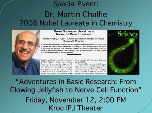
Chalfie to speak at Univ of San Diego, Nov. 12, 2010
|
|
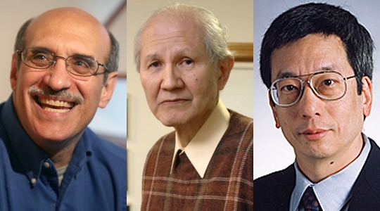
| Martin Chalfie, Osamu Shimomura, and Roger Tsien (L to R) photo from here |
 |
 |
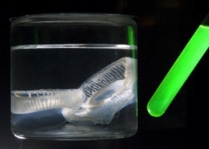 |
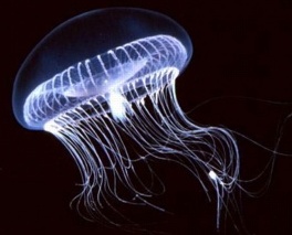
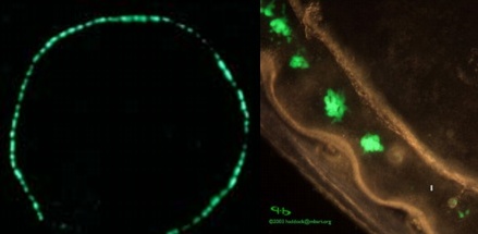
Aequorea victoria | Aequorea photo-organs fluorescing green [The GFP Site] | Aequorea and purified GFP [Woods Hole MBL] |
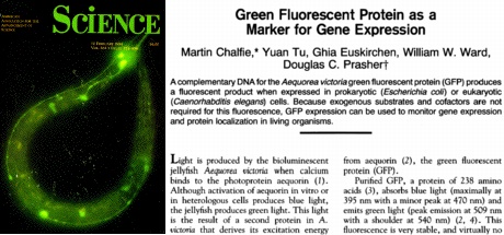
The gene for GFP was cloned at Woods Hole by Dr. Douglas Prasher in 1992. Martin Chalfie (Columbia University) had learned of Prasher's efforts, and recognized that GFP might make an excellent marker for cells. After some delay, Chalfie obtained the DNA for the GFP gene from Prasher, and fused the GFP gene to a worm (C. elegans) gene control region. The result was glowing cells in a live worm. Very soon, GFP and its derivatives were being used as glowing cellular markers in nearly every organism, tissue or cell under study in which such DNA manipulations can be made. The fluorescent proteins could also be used to see subcellular structures in many colors.
 |
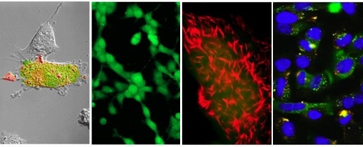 |
Chalfie published GFP results in the journal 'Science' in 1994 | Cells with various fluorescent markers [Tsien lab] |
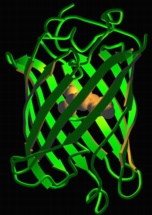
Like Chalfie, many scientists including Roger Tsien recognized the nearly limitless potential of GFP for research. Tsien and colleages (Univ of California, San Diego) studied the formation of the fluorescent molecule created when GFP folds into its characteristic 'beta barrel' structure, and subsequently generated many variants of GFP and other fluorescent proteins that make a rainbow of colors so a single live animal or tissue can be marked with several different colored proteins simultaneously. Other scientists have also studied and tinkered with GFP, or been inspired by this work to isolate and characterize fluorescent proteins from other organisms (such as corals).
 |
 |
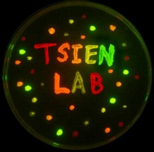 |
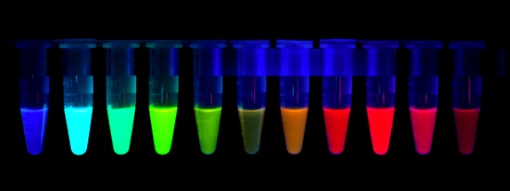
GFP structure [Tsien lab] | Rainbow of fluorescent proteins [Tsien lab] | Glowing bacteria on culture plate [Tsien lab] |
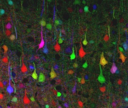
Perhaps the ultimate expression of color diversity using fluorescent proteins is the 'Brainbow' in which more than 100 different colors can be generated in different nerve cells of the brain, aiding study of nerve cell function and diversity. Fluorescent proteins have also been used for more whimsical purposes (or pernicious, depending on your point of view), such as making fluorescent aquarium fish (GloFish ®) .
 |
 |
 |
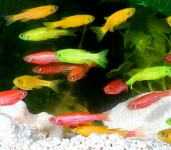 |
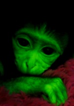
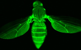
Rhesus monkey with GFP | Drsophila with GFP | The 'BrainBow' | 'GloFish ®' zebrafish (Danio) |
Shimomura:
Press release from the Marine Biological Laboratory at Woods Hole
Chalfie:
Marty Chalfie's Home Page at Columbia University, Dept of Biological Sciences
Tsien:
Roger Tsien's Lab Home Page at the Univ of California, San Diego