 Click for larger version
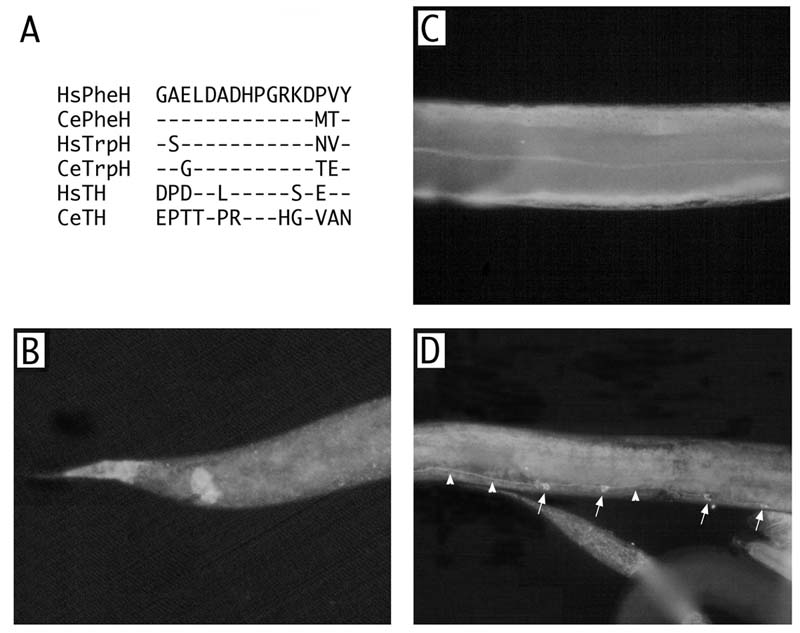
 Staining in C. elegans hypodermis and neurons with PH8 monoclonal antibody and TRITC-conjugated secondary antibody (goat anti-mouse IgG). (A) Alignments of protein sequences with the PH8 epitope. The top sequence is the chymotryptic fragment of human PheH recognized by PH8 (Cotton et al., 1988). The antibody recognizes vertebrate PheH in situ and on western blots, and recognizes vertebrate TrpH and TH in situ under appropriate conditions. Based on the sequence identity shown we expect the antibody to recognize at least C. elegans PheH (K08F8.4) and TrpH (ZK1290.2). (B) PH8 staining in larval tail. (C) Staining in adult body wall. Note intensity is greater in lateral hypodermal seam and ventral hypodermal cells than the rest of the hypodermis (D) Staining of male-specific serotonergic CP neuronal cell bodies and processes in the ventral nerve cord of an adult male. Four of six cell bodies can be seen (arrows). Processes in the ventral nerve cord are indicated with arrowheads.